On this page:
• The Structure of the Human Eye
• How Light is Regulated and Images are Brought into Focus
• The Retina and Function of Rods and Cones
• Rods and Cones in Seeing Colour and Distinguishing Form
Black Friday sale 8% off

• The Structure of the Human Eye
• How Light is Regulated and Images are Brought into Focus
• The Retina and Function of Rods and Cones
• Rods and Cones in Seeing Colour and Distinguishing Form
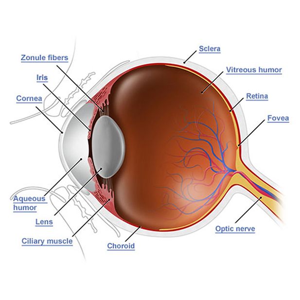
Please click on the links to find out about the different parts of the human eye.

The cornea is a clear, transparent area that covers the iris and the pupil. It allows light to enter the eye and then refracts light to the retina enabling both sight and focus. The cornea makes up two-thirds of the eye's total optical power.
The lens is embedded in the anterior segment of the eye and held in place by a ring of fibrous tissue, it is curved and transparent. Its purpose is to reflect light rays onto the retina at the back of the eye with the help of the cornea.
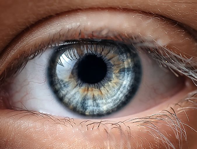
The iris, which is a coloured circular shaped and muscular structure, gauges the amount of light that will enter into the eye, or travel through to the pupil, which is positioned centrally inside of the iris. The pupil constantly adjusts opening and closing, depending on the amount of light coming through, or dimness of the setting. The opening and closing process of the pupil is medically known as dilation and miosis.
When light passes through to the pupil, it rests upon the lens, which is a translucent and supple structure, attached by connective tissue fibres to spherically arranged smooth muscle tissue. This muscle fibre is known as the ciliary muscle.
Is a layer of light-sensitive tissue which lines the inner surface of the eye, it is used to convert images received from the eyes lens to the optic nerve so the brain can process them.
The Aqueous humor is found between the lens and the cornea and is an area of clear fluid that provides nutrients. The fluid is produced in the ciliary body, and helps to protect the eye against dirt, dust and other pathogens that can be transported by wind towards the eye's surface.
Is a ring of smooth muscle in the middle layer of the eye that holds the lens in place, it also helps to maintain the eyes focus when viewing objects at different distances by assisting in the changing shape of the lens. Fluid from the aqueous humor is also regulated by the ciliary muscle.
The Zonule also referred to as the ciliary zonue is a ring of delicate fiberss connecting the eye's lens and with the ciliary body to keep everything in place. The zonule itself is split into two separate layers. One layer is thick and made from a group of fibres known as the zonular fibers, whereas the other layer is thinner and lines the hyaloid fossa and area in front of the eye's lens.
The sclera, which is the white part of the eye, is a resilient outer coating. The function of this structure is to protect the eyeball. The clear cornea is connected to the sclera and its role is vital in the process of vision. The cornea is responsible for bending light that passes through it, doing so in order to establish focus. As the light travels through the cornea, it is directed to the aqueous humor, which is a structure that contains fluid. The aqueous humor functions to provide nourishment and give cushioned support to the cornea and lens.
The main chamber of the eye is called the vitreous humor and it is here that the light then passes through, bathing the sides and the back of the eye, to fully cover the retina. This structure is primarily made up of photoreceptor cells, blood vessels and neurons.
The choroid is located in between the retina and the sclera and this structure is made up of blood vessels and pigmented cells that absorb light that has gone unrecognised by the photoreceptors. The action initiated by the choroid is very important, as it prevents the distortion of reflected light from occurring. The blood vessels are present to provide essential nourishment to the retina.
The Optic Nerve Located right at the back of the eyeball is the optic nerve, a structure responsible for transmitting information to the thalamus region of the brain, which is involved in the processing and relaying of sensory signals. The information received via the thalamus is then passed over to the visual cortex for analysis. Skeletal muscles enclose the eye, directing and assisting the movement of the eyeball, allowing you to look from left to right and up and down.
The optic disk is another structure found on the retina and is the place where the axons of the optic nerve exit the eye. The optic nerve is insensitive to light. In fact this is the area where your sight becomes obstructed. This is the reason why it is termed as being the blind spot.
The fovea, also known as the fovea centralis, is a tiny funnel shaped cavity that contains the highest amount of photoreceptors in the entire eye. This miniature structure is located directly on the retina and when you cast your gaze downwards, the image your glance is directed at is focused onto the fovea.
The iris is responsible for monitoring the amount of light that enters the eye, with the aide of two pairs of smooth muscle structures. In bright light, the muscle contracts around the pupil, causing the pupil to contract. If this process didn't occur, the bright and intense light would overpower your photoreceptors, causing temporary blindness. When light is dim, the smooth muscle, positioned around the pupil, contracts, causing the pupil to open wide and dilate.
The muscles are controlled by nerves, which is the reason why a physician shines a light into an injured or unconscious person's eye, to assess the condition of his or her nerves. When light is shone into the eye, pupils that are functioning properly should contract and when the light is removed the pupils should respond by dilating. Fixed or dilated pupils are often an indication of an extensive malfunction of the nervous system and should be taken very seriously. Certain drugs can also make the pupils dilate.
The light that comes into the eye focuses on both the cornea and the lens. The cornea has a curved shape and is the structure that bends most of the light that comes into the eye. The curved shape of the cornea is immobile and cannot be adjusted. The regulating capacity responsible for bending the light and the ability to modify focus between distant and close objects is achieved by an adjustment of the curved shape of the lens, which is referred to as being 'accommodation'.
The ciliary muscle adjusts the curvature of the lens with contractions and as the inner radius of the muscle shrinks, the amount of tension on the fibres that attach to the lens is reduced. This helps the lens to protrude outwards, which is necessary when focusing on an object positioned at close proximity. When the ciliary muscle is in a relaxed mode, an increase of tension occurs in the ring of muscle, which works to flatten and stretch it, pulling distant objects into focus.
The flexibility and suppleness of the lens is integral to sight and good vision and the condition presbyopia is illustrative of the changes that occur when the lens becomes more rigid and stiff with age, causing blurred and hazy vision. This condition eventually affects everyone to varying degrees and sets in after the age of 40. The stiffening of the lens restricts the bulging or protruding action and even when the ciliary muscles are contracted to their maximum degree, the protruding shape cannot take effect.
If you notice that you have to hold reading material further away from your eyes in order to focus more clearly and you are over 40, you may be experiencing presbyopia and should schedule an appointment with your optician for a diagnosis.
Eyesight is achieved when light is converted into impulses by the eye, a process which occurs inside of the retina. The distinctive formation of the retina enables you to see objects in full colour, have perception of various objects and adapt to different intensities of light. The retina is made up of four layers, consisted of the choroid, which operates to absorb the light that is not picked by photoreceptor cells.
The next layers of the retina are the rods and cones, which are the photoreceptor cells. They are called this because of their unique shape. These photoreceptors (rods and cones) meet with the third layer, which consists of neurons named bipolar cells; which are partially responsible for the integration of information, before passing it on to the next and fourth layer.
This layer is made up of ganglion cells, which are also neurons. Ganglion cells have long axons that become the optic nerve that leads to the brain. Even though light has to pass through the third and the fourth layer to get to the photoreceptor cells, this does not affect or limit the photoreceptor cells capability to receive and transform light.
The photoreceptor cells, which are the rods and cones, are quite intricate in their structure and function. At one end of the cell there is a succession of flat disks that are arranged to create a structure shaped as a rod or cone. Inside of the flat disks are multiple molecules made up of a protein called photopigment.
As soon as a photopigment receives light the shape of its molecules are altered, which causes the photoreceptor to shut down some sodium channels, in an attempt to reduce the number of neurotransmitters released.
Neurotransmitters, released by rods and cones, tend to slow down the bipolar cells; therefore when light is introduced the activity of bipolar cells will ultimately increase. This, as a consequence, activates the ganglion cells.
There are in the region of 120 million rods and 6 million cones in the retina. The cones and rods clearly outnumber the ganglion cells that consist of around 1 million.
Rods have great sensitivity to dim light and although rods help you to see better when the light is dim, they are not able to pick up on colour or make out intricate images. In fact they only provide vision in black and white. The part of the fovea that is furthest away from the retina is populated by the highest number of rods, in comparison to cones.
Cones are responsible for the colour in your vision and the detailing of images. This is due to the variations of coloured cones contained in the retina, which consist of blue, green and red selections. Each of the coloured cones contains a photopigment that can absorb the energy of their specific colour tones especially well.
Different colours are distinguishable because of the way in which the brain reads the percentage of impulses that come from the ganglion cells that connect to the different coloured cones. White light is perceived when all three cones are simultaneously activated by various wavelengths.